Gallery
phagocyte images: illustrations and micrographs. All credits and licenses listed.
Micrographs
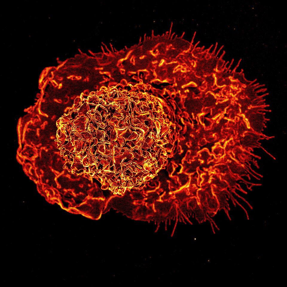
Scanning electron micrograph of a macrophage. The cell surface shows irregular ruffles and pseudopodial extensions characteristic of an activated macrophage.
NIAID (National Institute of Allergy and Infectious Diseases) · CC BY 2.0 · source
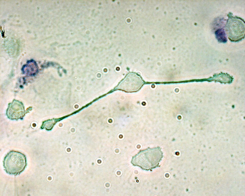
Light micrograph of a macrophage.
Obli (original uploader, English Wikipedia) · CC BY-SA 2.0 · source
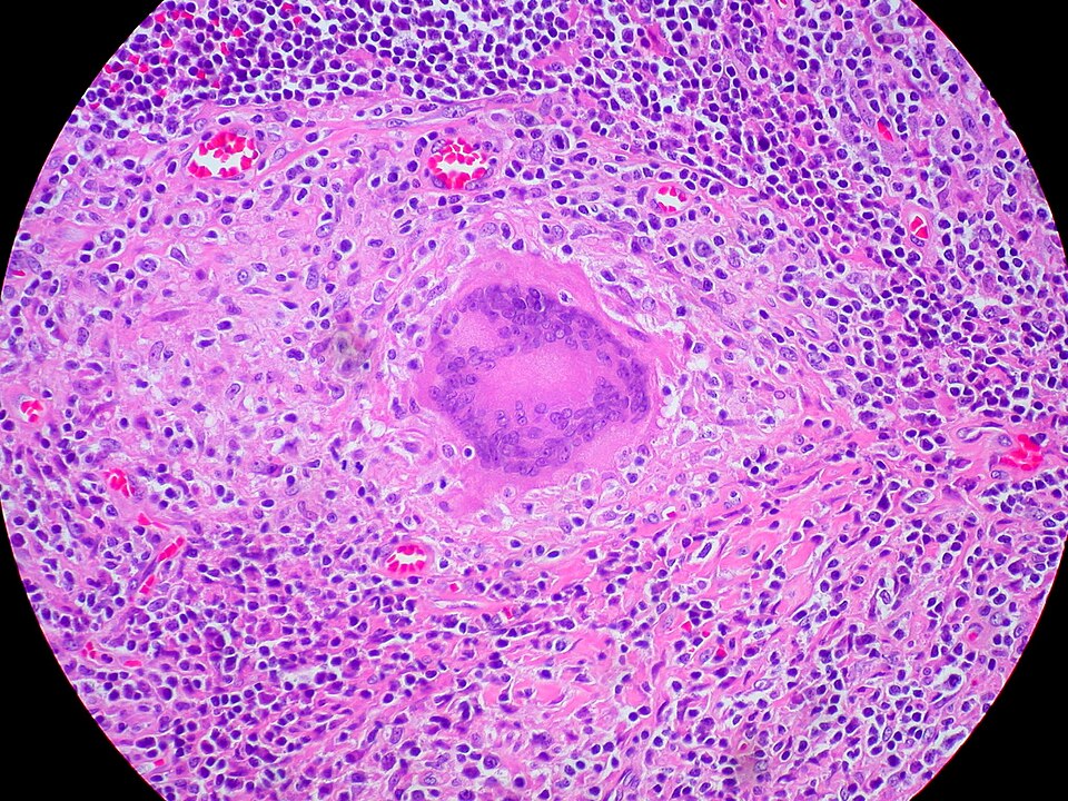
Langhans giant cell in a caseating tuberculous granuloma. Haematoxylin and eosin. Human tissue. The peripheral arrangement of nuclei is characteristic of Langhans type, distinguishing it from the scattered nuclei of foreign-body giant cells.
Ed Uthman, MD (from Houston, TX, USA) · CC BY 2.0 · source
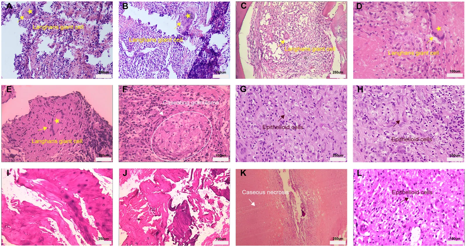
Representative H&E histopathology of spinal tuberculosis lesions showing recurrent pathological patterns. Yellow arrows and asterisks: multinucleated Langhans-type giant cells. White arrows and circles: caseous granulomas. Brown-red arrows: epithelioid cells. Human tissue.
Liu, Guo, Liu et al. 2026, Frontiers in Immunology (PMC13541710) · CC BY 4.0 · source
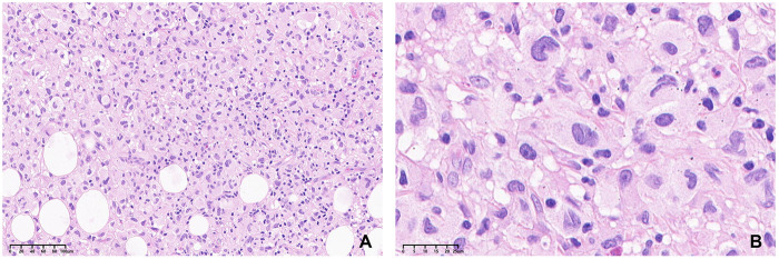
Pathology from the initial biopsy. The lesion consisted of large neoplastic cells with abundant eosinophilic cytoplasm, eccentrically placed nuclei, and increased mitotic activity. (A) H&E stain (X200), (B) H&E stain (X800).
Li M, Hu T, Liu Z, Yu H et al. 2026, Frontiers in Surgery (PMC13478121) · CC BY 4.0 · source
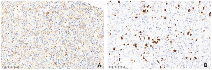
Immunohistochemical analysis of histiocytic sarcoma. (A) Neoplastic cells positive for CD68 (IHC×200), (B) Ki-67 labeling index was about 30% (IHC×200).
Li M, Hu T, Liu Z, Yu H et al. 2026, Frontiers in Surgery (PMC13478121) · CC BY 4.0 · source
Illustrations
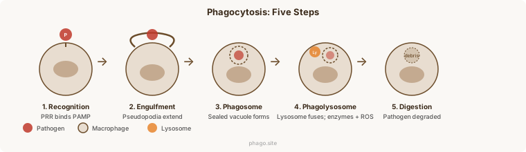
Phagocytosis: five steps from recognition to digestion. Labeled illustration.
phago.site · original
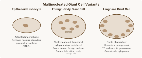
Multinucleated giant cell variants: epithelioid histiocyte, foreign-body type (scattered nuclei), and Langhans type (peripheral nuclei). Labeled illustration.
phago.site · original
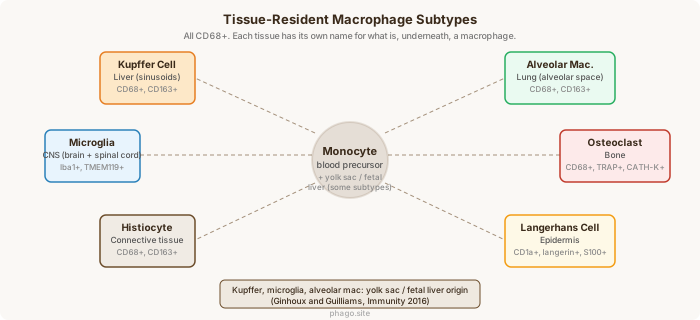
Tissue-resident macrophage subtypes: six named variants with tissue location and key markers.
phago.site · original